|
Bagus Printer Shop
|
GE Vivid i
|
|
|
| Add to My Favorites | |
| HiSupplier Escrow |
Product Detail
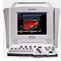
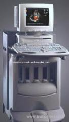
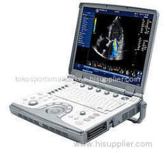
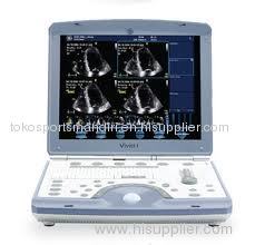
GE Vivid i
GE Healthcare's new Vivid i is the world's first miniaturized cardiovascular ultrasound system to provide high-performance, full-featured imaging in a lightweight design.
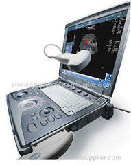
The GE Vivid i addresses one of the biggest challenges in cardiovascular care – access to complete, real-time diagnostic information. The new system expands the reach of echocardiography by offering all the functionality and high performance of full-featured premium scale systems – but in a completely portable and wireless design that weighs 30 times less.
GE developed Vivid i by miniaturizing the components of a premium echocardiography system weighing more than 180 kilograms to provide a portable system weighing less than five kilograms. Vivid i features wireless functionality, enabling physicians to transfer files instantly from the system to other physicians for consultation, or to the patient’s bedside so that they can be more informed and involved in their healthcare decisions.
We offer big savings over the cost of purchasing a new ultrasound machine.
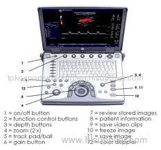
Vivid i Features:
- Phased-array transducer technology for 2D, color and Doppler imaging.
- Multiple focal zones help optimize image quality.
- Extremely high frame rates, enhanced color flow and color angio assist in acquiring extremely low- velocity flows.
- 5 levels of Coded Octave Harmonics help you obtain quality images from difficult-to-image patients.
- LVO contrast option, based on 2D Harmonic imaging with Coded Phase Inversion, aids in scanning your most challenging patients.
- Triplex and duplex display capabilities to simplify Doppler acquisitions.
- Automatic Tissue Optimization (ATO) option helps you obtain quality images faster by automatically adjusting the image settings to optimize images.
- Complete measurement and analysis worksheets and reports are tailored to each exam.
- Live Anatomical M-Mode corrects for off-axis orientation in situations where the heart is not positioned or shaped normally.
- Tissue Doppler Imaging provides real-time Doppler spectral information for specified myocardial motion, allowing for instantaneous tissue velocity measurements.
- Over 40 gigabyte disk space for archive storage of patient
- Full DICOM connectivity with embedded raw data speeds exams by allowing you to perform post-exam quantitative analyses at your convenience on the integrated EchoPAC or optional EchoPAC Dimension workstation.
- MPEGvue option compresses images into compact file sizes. These high quality images can be transferred to any compatible media or network, or e-mailed directly for viewing.
- eVue option permits remote interactive monitoring of images on any PC, via wired or wireless network communication, for fast and convenient consultations.
- Lightweight, 5 kg package makes system transport fast and easy – simply strap on your back or carry to your patients.
- Ultra-small footprint reduces operational costs by eliminating the need for special rooms, lifts or vans.
- Rechargeable battery provides up to 1.0 hour of full scan operation
Applications: Cardiac, Vascular
Related Search
Find more related products in following catalogs on Hisupplier.com
Company Info
Bagus Printer Shop [Indonesia]
Business Type:Trading Company, Distributor/Wholesaler
Country/Region: Indonesia


.gif)